Sessions & Tracks
Track 1. Comprehensive Eye Care:
At the point when an individual calls to make an eye appointment, the person ought to be set up to portray any present vision issues. What's more, patients ought to inquire as to whether the eye examination will influence their vision briefly and in the event that they will require somebody to drive them home. They may likewise need to get some information about the expense of the test, if their protection plan will take care of any of the expense, and how installment is taken care of.
Prior to setting off to the arrangement, patients should assemble the accompanying data to help answer addresses the eye care proficient may inquire:
Side effects of current eye issues (flashes of light, trouble seeing around evening time, brief twofold vision, loss of vision, and so forth.).
Eye wounds or eye medical procedures (estimated dates, where treated)
Family ancestry of eye issues (glaucoma, macular degeneration, waterfalls, and so on.)
Any inquiries regarding their vision, glasses, contacts, laser medical procedure, and so forth.
A rundown all things considered and over-the-counter medications right now being utilized
Their general wellbeing condition (hypersensitivities, unending medical issues, activities, and so on.)
Patients ought to likewise convey the accompanying things with them to their eye arrangement
Glasses, contact focal points or both
A rundown all things considered and over-the-counter medications as of now being taken
Restorative or medical coverage card
Personal ID
Development Of Eye
Orbit
Eyelids
Glandular Apparatus
Extra-Ocular Muscles
General Ophthalmology
Clinical Ophthalmology
Track 2. Smartphones and Eyestrain:
As much as we rely upon our cell phones for survey and reacting to messages, checking the climate, perusing feature news, and posting notices on Facebook, our cell phones might cause us some vision issues. Gazing at those minor screens can expedite a variety of eye issues, for example, obscured vision, migraines, sore eyes, cerebral pains, muscle strain and dry eye.
Cell phones can cause different issues too. The iPhone's most current update appears to influence offset and soundness with the new symbols zooming in and out. Clients have griped of discombobulation. Perusing in bed can influence rest designs because of the blue light radiated from the screen. This light can diminish dimensions of melatonin and make it harder to nod off. For all the assistance that our telephones and electronic gadgets offer, they are actually giving us a migraine.
The appropriate response isn't to quit utilizing your cell phone. Or maybe, actualize a framework where you take customary breaks about at regular intervals or thereabouts. This is known as the 20-20-20 rule. Like clockwork, gaze something like 20 feet away for no less than 20 seconds. This will help rest your eyes and forestall weariness and strain that causes those commonplace cerebral pains, soreness and obscured vision. This is particularly vital for kids who might be new to having a telephone or who may not make sure to give their eyes a rest. Try not to be hesitant to define some reasonable limits and rules for youngsters so they can learn discretion and control in utilizing their electronic gadgets.
Visual Snow
20-20-20 Rule
Colour Temperature
Reading related disorder
Computer Eyewear
Track 3. Latest Research In Ophthalmology And Vision Improvement:
Development is characterized as the presentation of something new. Ophthalmology and its sub-claims to fame have been at the bleeding edge of therapeutic development and have grasped the fast advances in different innovations, including pharmacology, imaging, information handling, and gadgets. In spite of the fact that the beginnings of a significant number of these advances started before, this year saw them start to grab hold by ophthalmologists and other eye care experts, with a definitive objective of improving the personal satisfaction for our patients. 8 Key Technical Advances in Ophthalmology are Cataract surgery techniques, Equipment to improve accuracy, New glaucoma shunts, Advances in ocular imaging, Implants for macular degeneration, EMR systems for ophthalmology practices, Premium IOLs, Femtosecond laser for cataract surgery
3-D Retina Organoid Challenge
Translational Research
eyeGENE
Regenerating The Optic Nerve
Bionic Eyes
Optical Coherence Tomography
Robot Surgery
Lens Technology
Optical Lens and Processing System
Track 4. Retina And Retinal Disorders:
Retinal maladies change broadly, yet the greater part of them cause visual manifestations. Retinal illnesses can influence any piece of your retina, a slight layer of tissue within back mass of your eye. The retina contains a huge number of light-delicate cells (poles and cones) and other nerve cells that get and sort out visual data. Your retina sends this data to your mind through your optic nerve, empowering you to see. Treatment is accessible for some retinal infections. Contingent upon your condition, treatment objectives might be to stop or moderate the ailment and safeguard, improve or re-establish your vision. Untreated, some retinal infections can cause serious vision misfortune or visual deficiency.
Floaters
Macular Degeneration
Diabetic Eye Disease
Retinal Detachment
Retinitis Pigmentosa
Retina Vein Impediment
Cone-Rod Dystrophy
Retinal Artery Occlusion
Acquired Retinal Issue
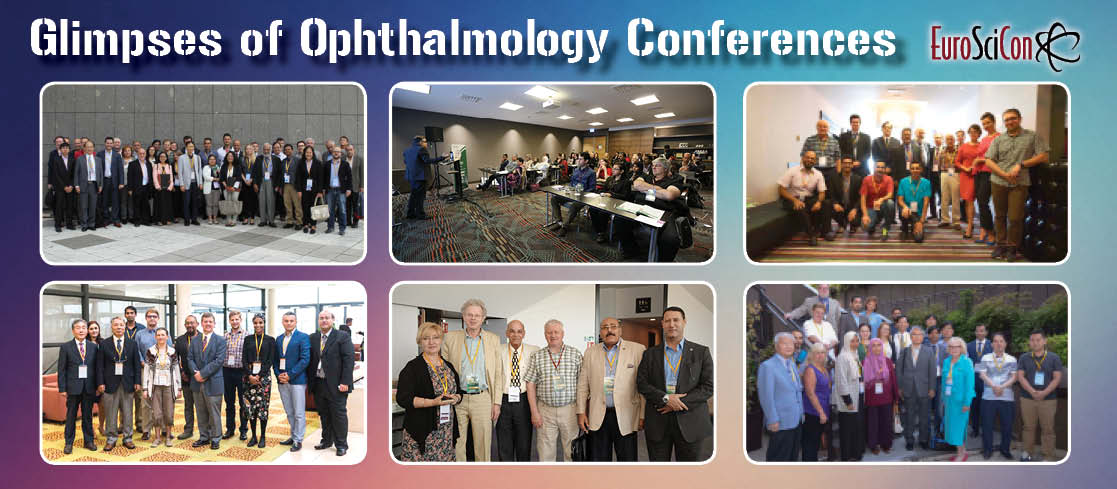
Conference Image
Track 5. Cornea and Corneal Diseases:
The cornea is the eye's peripheral layer. It is the unmistakable, domeshaped surface that covers the front of the eye. It assumes a vital job in centering your vision. In spite of the fact that the cornea may look clear and appear to need substance, it is an exceptionally composed tissue. In contrast to most tissues in the body, the cornea contains no veins to feed or secure it against contamination. Rather, the cornea gets its sustenance from tears and the watery silliness (a liquid in the front piece of the eye that lies behind the cornea).The cornea goes about as a hindrance against earth, germs, and different particles that can hurt the eye. The cornea imparts this defensive undertaking to the eyelids and eye attachments, tears, and the sclera (white piece of the eye). The cornea additionally assumes a key job in vision by helping center the light that comes into the eye. The cornea is in charge of 65-75 per cent of the eye's all out centering power.
The cornea goes about as a boundary against soil, germs, and different particles that can hurt the eye. The cornea imparts this defensive errand to the eyelids and eye attachments, tears, and the sclera (white piece of the eye). The cornea additionally assumes a key job in vision by helping center the light that comes into the eye. The cornea is in charge of 65-75 per cent of the eye's absolute centering power. The cornea and focal point of the eye are worked to concentrate light on the retina, which is the light-delicate tissue at the back of the eye. At the point when light strikes the cornea, it twists—or refracts—the approaching light onto the focal point. The focal point refocuses that light onto the retina, which begins the interpretation of light into vision. The retina changes over light into electrical driving forces that movement through the optic nerve to the cerebrum, which deciphers them as images. The refractive procedure the eye utilizes is like the manner in which a camera snaps a photo. The cornea and focal point in the eye go about as the camera focal point. The retina resembles the film (in more seasoned cameras), or the picture sensor (in computerized cameras). In the event that the picture isn't centered legitimately, the retina makes a hazy picture. The cornea likewise fills in as a channel that screens out harming bright (UV) light from the sun. Without this security, the focal point and the retina would be presented to damage from UV beams.
Corneal Transplant
Corneal Abrasions
Corneal Innervation
Corneal Collagen Cross-Linking
Synthetic Corneas
Penetrating Keratoplasty
Lattice Dystrophy
Fuchs’ Dystrophy
Map Dot Fingerprint Dystrophy
Track 6. Glaucoma:
Glaucoma is a condition that makes harm your eye's optic nerve and deteriorates after some time. It's frequently connected to a development of weight inside your eye. Glaucoma will in general be acquired and may not appear until some other time throughout everyday life. The expanded weight, called intraocular weight, can harm the optic nerve, which transmits pictures to your cerebrum. On the off chance that the harm proceeds, glaucoma can prompt perpetual vision misfortune. Without treatment, glaucoma can cause all out lasting visual impairment inside a couple of years. A great many people with glaucoma have no early side effects or agony. You have to see your eye specialist consistently so she can analyse and treat glaucoma a little while later term visual misfortune occurs. In case you're over age 40 and have a family ancestry of the infection, you ought to get a total eye test from an eye specialist each 1 to 2 years. In the event that you have medical issues like diabetes or a family ancestry of glaucoma or are in danger for other eye sicknesses, you may need to go all the more frequently.
Cyclophotocoagulation
Iridotomy
Trabeculoplasty
Primary And Secondary Glaucoma
Open And Closed Angle Glaucoma
Medical Therapy Of Glaucoma
Pigmentary Glaucoma
Steroid Induced Glaucoma
Inflammatory Glaucoma
Medical Therapy Of Glaucoma
Track 7. Cataract And Refractive Surgery:
Refractive medical procedure is a strategy for remedying or improving your vision. There are different surgeries for revising or changing your eye's centering capacity by reshaping the cornea, or clear, round arch at the front of your eye. Different techniques include embedding a focal point inside your eye. The most broadly performed sort of refractive medical procedure is LASIK (laser-aided situ keratomileusis), where a laser is utilized to reshape the cornea. For individuals who are astigmatic, certain refractive medical procedure methods will decrease the shape of a cornea that is excessively steep with the goal that the eye's centering power is diminished. Pictures that are engaged before the retina, because of a more extended eye or soak corneal bend, are driven nearer to or straightforwardly onto the retina following medical procedure. Farsighted individuals will have refractive medical procedure methods that accomplish a more extreme cornea to build the eye's centering power. Pictures that are engaged past the retina, because of a short eye or level cornea, will be destroyed nearer to or legitimately onto the retina after medical procedure. Astigmatism can be rectified with refractive medical procedure methods that specifically reshape bits of an unpredictable cornea to make it smooth and symmetrical. The outcome is that pictures center obviously around the retina instead of being contorted because of light dissipating through an unpredictably molded cornea.
Refractive medical procedure may be a decent alternative for you in the event that you:
Need to diminish your reliance on glasses or contact focal points;
Are free of eye ailment
Acknowledge the inalienable dangers and potential reactions of the technique;
Comprehend that you could even now need glasses or contacts after the method to accomplish your best vision;
Have a proper refractive blunder.
There is no generally acknowledged, best strategy for remedying refractive blunders. The best choice for you ought to be chosen after an intensive examination and exchange with your ophthalmologist. On the off chance that you are thinking about refractive medical procedure, you and your ophthalmologist can talk about your way of life and vision needs to decide the most proper method for you.
Secondary Cataract
Traumatic Cataract
Congenital Cataract
Radiation Cataract
Nuclear Cataract
Cortical Cataract
Posterior Sub-Capsular Cataract
Congenital Cataract
Visual Acuity Test
Dilated Eye Exam
Tonometry
Track 8. Dry eye and Uveitis:
Uveitis is an expansive term for some issues with your eye. What they share for all intents and purpose is eye irritation and swelling that can annihilate eye tissues. That annihilation can prompt poor vision or visual deficiency. "Uveitis" is utilized in light of the fact that the swelling regularly influences the piece of your eye called the uvea. Your eye is made of layers. The uvea is the center layer. It's between the white piece of your eye called the sclera and the internal layers of your eye.
Your uvea contains three essential structures:
The iris. That is the shaded hover at the front of your eye.
The ciliary body. Its main responsibility is to enable your focal point to center and make the liquid that supports within your eye.
The choroid. This is a gathering of veins that give your retina the supplements it needs.
Are There Different Types of Uveitis?
Indeed. Which type you have relies upon where the swelling is.
Front uveitis is the most well-known. It influences the front of your eye.
Halfway uveitis influences your ciliary body.
Back uveitis influences the back of your eye.
In the event that your whole uvea is excited, you have pan uveitis.
Dry Eye or Keratoconjunctivitis sicca disorder (KCS or dry eye) is an issue of major epidemiologic significance. It influences truly a great many individuals around the world, with ladies significantly over spoke to, especially those ladies who have entered menopause. The issue may go with dry mouth, and might be found in relationship with a fundamental malady, for example, rheumatoid joint pain or foundational lupus erythematous. It is, in numerous occasions, definitely in excess of a basic "aggravation" issue. It has the potential for genuine visual outcomes, starting with the arrangement of dry spots on the cornea, advancing to epithelial deformities or "scraped spots" which oppose mending, and after that in certain occasions eventuating to ulceration of the cornea, once in a while even with blunt perforation. The utilization of mitigating and immunomodulatory specialists by topical application additionally assume a job in numerous patients' dry eye care, since irritation of the lacrimal organ has been appeared to be available and to obstruct the ordinary capacity of that tear-creating organ in a huge extent of patients with dry eye ailment. Subsequently, topical Alrex (a feeble corticorsteroid) and topical Restasis (containing cyclosporin) have been appeared to be helpful in such patients. Xiidra (Lifitegrast) is another safe arbiter dropfor the board of dry eye appeared to be viable too. Moreover, dietary supplementation with nourishments wealthy in omega 3 free unsaturated fats, for example, chilly water fish (eg, salmon) can likewise help in the rebuilding of the typical wellbeing and working of the broken lacrimal and Meibomian organs. Lipiflow methodology is another alternative for treatment of Meibomian organ brokenness and Blepharitis. At long last, the utilization of extraordinarily built scleral focal points can moreover be useful in holding a layer of fluid against the cornea in patients who have significant absence of satisfactory tears.
Heavy eyelids
Eye fatigue
Stinging or burning
Kerato-conjunctivitis sicca
Dysfunctional tear syndrome
Anterior uveitis
Intermediate uveitis
Intermediate uveitis
Posterior uveitis
Diffuse uveitis (also called pan uveitis)
Track 9. Ocular Trauma:
Visual injury is a standout amongst the most under-perceived reasons for vision misfortune in the created world. Obtuse or infiltrating visual injury can prompt vision misfortune through waterfall or glaucoma. Etiologies of visual damage contrast in urban regions contrasted with different settings, and vary from nation to nation, between various districts of the world, and between varying statistic or financial classes. Epidemiological examinations can experience trouble because of here and there remote injury and trouble getting an exact history. Techniques for avoidance of visual injury require learning of the reason or system of damage, which may empower progressively fitting focusing of assets toward anticipating such wounds. Both eye injury exploited people and society bear a vast, possibly preventable weight.
General and complete assessment of a harmed patient and a harmed eye is critical to spare life and sight. Suitable absence of pain, sedation, and security of open globe from further damage are an imperative piece of the assessment. Enemies of emetics ought to be managed to maintain a strategic distance from Valsalva move if the patient has queasiness or regurgitating to avoid conceivable expulsion of intraocular substance.
1. Vital signs ought to be checked constantly. Capture of an intraocular muscle may prompt hazardous bradycardia, especially in kids, along these lines pulse ought to be observed. Brief appraisal of other hard and delicate tissue wounds must be performed. When the patient is observed to be steady, ophthalmic assessment can continue securely.
2. Introductory visual examination The ophthalmologist ought to have a high file of doubt for harm to different pieces of the eye when horrible waterfall as well as glaucoma are available. Worry for harm to the foremost chamber point structures, zonular strands, corneal endothelium, vitreous, and retina ought to be high. Consideration ought to be paid to the trustworthiness of the focal point case.
3. Visual Acuity: Important indicator for the visual injury score, Vision ought to be tried with amendment and pinhole utilizing Snellen's/ETDRS outline. In the event that vision is lower than this, finger checking might be recorded; if this is preposterous, hand movement or light discernment might be recorded. No light observation is a vital thought, as eyes with this dimension of vision may demonstrate some improvement following appropriate investigation and the executives.
4. Pupillary response: Relative afferent pupillary imperfection is an imperative negative prognostic factor for influenced eye. This finding should raise doubt for optic nerve harm, separation, and compartment disorder if different signs are available.
5. Intraocular Pressure: IOP might be high, low or typical after damage. Low intraocular weight or hyoptony should raise alert amid test for cracked globe, and care ought to be taken to shield the eye from expulsion of substance. The eye ought to be protected when not under examination.
6. Motility: It is imperative to evaluate visual motility as there are odds of immediate or backhanded harm to additional visual muscles. Extraocular muscle capture must be precluded.
7. Field Testing: Central field can be assessed by the Amsler network and fringe field by encounter strategy, in any case, this is, best case scenario a gross assessment and ought to be followed up via mechanized perimetry when conceivable/accessible.
Treatment incorporates Imaging, Electrophysiology, Laboratory tests, Medical treatment, Surgery, Surgical follow-up and so on.
Blunt Trauma
Ruptured eye heal
Open globe injury
Conjunctival chemosis
Retro bulbar Haemorrhage
Traumatic Optic Neuropathy
White-Eyed & Other Blowout Orbital Fractures
Lid Lacerations
Chemical Burns
Track 10. Diabetic Retinopathy:
Diabetic retinopathy happens when changes in blood glucose levels cause changes in retinal veins. Now and again, these vessels will swell up (macular oedema) and release liquid into the back of the eye. In different cases, unusual veins will develop on the outside of the retina. Except if treated, diabetic retinopathy can slowly turn out to be progressively genuine and advance from 'foundation retinopathy' to truly influencing vision and can prompt visual impairment. In the same way as other states of this nature, the beginning periods of diabetic retinopathy may happen without indications and without agony. A real effect on the vision won't happen until the illness progresses. Macular oedema can result from maculopathy and influence vision happens if releasing liquid causes the macular to swell. New vessels on the retina can incite dying, which can likewise square vision at times. Side effects may just end up perceptible once the ailment propels, yet the run of the mill indications of retinopathy to pay special mind to include:
Unexpected changes in vision/obscured vision
Eye floaters and spots
Twofold vision
Eye torment
Diabetic retinopathy is brought about by delayed high blood glucose levels. After some time, high sugar glucose levels can debilitate and harm the little veins inside the retina. This may cause haemorrhages, exudates and notwithstanding swelling of the retina. This at that point keeps the retina from oxygen, and anomalous vessels may develop. Great blood glucose control brings down diabetes retinopathy dangers.
Long haul great blood glucose level administration avoids diabetes retinopathy and lower the danger of creating it. Coronary illness chance factors additionally influence retinopathy hazard and incorporate ceasing smoking, having customary circulatory strain and cholesterol checks and experiencing normal eye registration.
The danger of creating diabetic retinopathy can be decreased through avoiding potential risk:
Taking an expanded eye examination once every year
Overseeing diabetes carefully through drug, insulin, diet and exercise
Test glucose levels routinely
Test pee for ketone levels routinely
Early Diabetic Retinopathy
Advanced Diabetic Retinopathy
Spots Or Dark Strings Floating In Your Vision
Impaired Colour Vision
Vitreous Haemorrhage
Track 11. Oculoplastics and Orbit Surgery:
Eye medical procedure, generally called visual medical procedure, will be medical procedure performed on the eye or its adnexa, customarily by an ophthalmologist. The eye is a fragile organ, and requires incredible consideration some time as of late, in the midst of, and after a careful strategy. An eye pro is accountable for picking the fitting careful strategy for the patient, and for taking the critical security shields. Notification of eye medical procedure can be found in a couple of out of date works. Today it continues being a comprehensively cleaned sort of medical procedure, having made distinctive techniques for treating eye issues. Since the eye is vivaciously given by nerves, anesthesia is fundamental. Neighborhood anesthesia is most usually used. Topical anesthesia using lidocaine topical gels are consistently used for quick techniques. Since topical anesthesia requires cooperation from the patient, general anesthesia is consistently used for adolescents, unpleasant eye wounds, and major orbitotomies and for disturbed patients. The specialist controlling anesthesia, or a restorative guardian anesthetist or anesthetist associate with capacity in anesthesia of the eye, screens the patient's cardiovascular status. Clean security measures are taken to set up the zone for medical procedure and lower the infection. These protections fuse the use of antiseptics, for instance, povidone-iodine, and sterile window hangings, outfits and gloves.
Eyelid Reconstruction
Radiotherapy
Chemosurgery
Lacrimal Surgery
C-DCR Conjunctivo-Dacryocystorhinostomy (tear duct bypass with implant)
Punctal Stenosis and Stent
Dacryocystorhinostomy Surgery
Cosmetic orbital surgery
Orbital decompression
Track 12. Ophthalmic Genetics:
Ophthalmic genetics is concerned with the genetic contribution to ophthalmic disease, including determination of patterns and risks of inheritance, as well as diagnosis, prognosis, and development of treatments for genetic abnormalities. . An extraordinary number of clutters with ophthalmic appearances are transmitted in trademark inherited examples through numerous ages, for the most part being owing to erasures, transformations, as well as duplications of little sections of explicit chromosomes of atomic DNA or the round DNA of mitochondria. Autosomal overwhelming issue incorporate neurofibromatosis type 1, tuberous sclerosis, Best vitelliform macular dystrophy, von Hippel– Lindau sickness, autosomal predominant optic decay, most instances of multifocal retinoblastoma, and a few instances of retinitis pigmentosa. Autosomal passive issue incorporate oculocutaenous albinism, rotate decay, xeroderma pigmentosa, and a few instances of retinitis pigmentosa. X-connected passive issue incorporate red-green partial blindness, X-connected retinoschisis, visual albinism, Norrie ailment, a few instances of retinitis pigmentosa, and most instances of choroideremia. Matrilineal legacy is normal for variations from the norm of mitochondrial DNA, for example, the point transformations that reason Leber's inherited optic neuropathy (LHON). Other mitochondrial issue, in which the trademark ophthalmic appearances are unending dynamic outside ophthalmoplegia (CPEO) and pigmentary retinopathy, may likewise be brought about by point transformations of mitochondrial DNA, yet in addition might be brought about by huge erasures of mitochondrial DNA, for example, in the Kearns– Sayre disorder, or changes of atomic DNA causing variations from the norm of mitochondrial work and acquired with an autosomal overwhelming or autosomal latent example. In many clutters with ophthalmic indications that obviously have a hereditary premise yet are once in a while transmitted through more than one age, the hereditary variation from the norm is a noteworthy or complete misfortune or duplication of at least one chromosomes including various qualities. Because of the nonattendance of a large portion of the typical supplement of qualities related with a specific chromosome in cases with complete chromosomal cancellations and to the nearness of half more than the ordinary supplement of qualities related with a specific chromosome in cases with complete chromosomal duplications, influenced people naturally have various morphological anomalies, as often as possible provoking chromosomal examination amid earliest stages or early youth. They are often sterile or ineffective in repeating. Much of the time, the strange supplement of chromosomes can be recognized by karyotyping.
Inheritance patterns
Genetic testing
Bardet-Biedl syndrome
Choroideremia
Stargardt disease
Achromatopsia
Juvenile X-linked retinoschisis
Colour Blindness
Retinitis pigmentosa
Anophthalmia and Microphthalmia
Polymorphism
Apoptosis
Crystallin
Optic Atrophy
Track 13. Neuro-Ophthalmology:
Neuro-Ophthalmology is the claim to fame that is worried about visual side effects coming about because of mind illnesses. The visual side effects can be separated into visual misfortune, or issues with eye developments. Visual misfortune may result from issues inside the optic nerve or its associations with the visual bits of the mind. The eye resembles a camera, and pictures are centered around the retina. The optic nerve is the link that takes these pictures to the mind. There are a few pieces of the cerebrum that are included with exact control of eye developments. Trouble inside these areas frequently creates misalignment of the eyes with resultant twofold vision. Any of a few procedures may influence the optic nerve or its associations; including strokes, irritation, various sclerosis or tumors. Visual misfortune may result from an issue inside the eye or its associations with the cerebrum. Obscured vision in one eye might be the consequence of a focal point issue (glasses), waterfall or different opacities obstructing light's passageway, a retinal issue, optic nerve illness or a sluggish eye (amblyopia). Twofold vision might be separated into two general classifications: monocular or binocular. Monocular twofold vision influences one eye at any given moment. In the event that the unaffected eye is shut, the twofold vision perseveres. Monocular twofold vision results from eye illness, for the most part a focal point issue or waterfall changes. Binocular twofold vision is possibly present when the two eyes are open and leaves when either eye is shut. Binocular twofold vision results from misalignment of the eyes. At the point when the eyes are pointed in various ways, the cerebrum sees two separate pictures (one from each eye).
The eyes are kept adjusted by the muscles that control eye developments, which thusly are constrained by three nerves from the cerebrum. Misalignment (causing binocular twofold vision) is most regularly because of an issue with one of three regions: the nerves that interface with the eye muscles, the association between the nerves and the eye muscles, or the eye muscles themselves. There are a few things that can influence the nerves setting off to the eye muscles including strokes or tumors pushing on the nerves. The most widely recognized malady to influence the association between the nerves and eye muscles is myasthenia gravis. There are a few muscle maladies that may influence the eye muscles.
Giant Cell Arteritis
Bell’s Palsy
Multiple Sclerosis
Headache
Optic Neuritis
Stroke
Diplopia (‘Double Vision’)
Internuclear Ophthalmoplegia
Neuro-Optometry
Neurology
Anisocoria
Ptosis
Nystagmus
Blepharospasm
Track 14. Paediatric Ophthalmology:
Kids are not grown-ups. They can't generally say what is pestering them. They can't generally respond to medicinal inquiries, and are not constantly ready to be patient and agreeable amid a therapeutic examination. Paediatric ophthalmologists realize how to look at and treat youngsters such that makes them loose and agreeable. Likewise, Paediatric ophthalmologists use hardware uncommonly intended for kids. Most Paediatric ophthalmologists' workplaces are orchestrated and enlivened in light of kids. This incorporates the examination rooms and lounge areas, which may have toys, recordings, and perusing materials for kids. This makes an agreeable and nonthreatening condition for your youngster. Paediatric ophthalmology is a sub-claim to fame of ophthalmology worried about eye sicknesses, visual advancement, and vision care in kids. Paediatric ophthalmologists can analyze, treat, and deal with all kids' eye issues.
Paediatric ophthalmologists by and large give the accompanying administrations:
Eye tests
Perform medical procedure, microsurgery, and laser medical procedure (for issues like powerless eye muscles, crossed eyes, meandering eyes, blocked tear pipes, retinal issues, and diseases)
Analyze issues of the eye brought about by illnesses of the body, for example, diabetes or adolescent rheumatoid joint inflammation (JRA) and other restorative and neurological ailments
Analyze visual handling issue
Care for eye wounds
Recommend eyeglasses and contact focal points
Child Abuse can make damage any piece of the eye. The most well-known indications are retinal haemorrhages (RHs) in babies and youthful youngsters with abusive head trauma (AHT). In spite of the fact that RHs are an essential marker of conceivable AHT, they are additionally found in different conditions. Recognizing the number, type, area, and example of RHs is essential in assessing a differential analysis. Eye injury can be found in instances of physical maltreatment or AHT and may incite referral for ophthalmologic evaluation. Doctors have a duty to think about maltreatment in the differential finding of Paediatric eye injury. Recognizable proof and documentation of dispensed visual injury requires an intensive examination by an ophthalmologist, including aberrant ophthalmoscopy, most ideally through a widened student, particularly for the assessment of conceivable RHs. An eye examination is useful in recognizing variations from the norm that can help distinguish a medicinal or awful etiology for beforehand well youthful youngsters who experience surprising and unexplained mental status changes with no undeniable reason, kids with head injury that outcomes in noteworthy intracranial discharge and cerebrum damage, and kids with unexplained demise.
Childhood eye misalignments and disorders
Strabismus
Paediatric glaucoma & cataracts
Retinopathy of prematurity
Shaken baby syndrome
Congenital birth defects
Blocked tear ducts
Ptosis
Nystagmus
Visual inattention
Paediatric cataracts
Paediatric glaucoma
Dyslexia and attention deficit disorder
Paediatric Ocular Surgery
Paediatric Neuro-Ophthalmology
Paediatric Optometry and Research
Track 15. Ocular Oncology and Ocular Pharmacology:
Tumor can make in a couple of structures in the eye zone and it happens when strong cells in or around the eye change and become uncontrollably, molding a mass called a tumor. A tumor can be good or ruinous. A positive tumor infers the tumor can grow however it won't spread. A destructive tumor is hazardous, which implies it can create and spread to various pieces of the body and development that structures in the eyeball is called an intraocular (inside the eye) risk. This fuses sorts like retinoblastoma, which is the ordinary sort of development affecting the eyeball in adolescents, Ophthalmologic infection, in which in case it impacts interior piece of the eye, those affecting the outside of the eye are extra visual. Ophthalmologic Tumors can be managed by chemotherapy; drug can be injected into the eye, making this treatment extraordinarily important for developments that have spread. Chemotherapy can be significant for treating intraocular (eye) lymphoma, and it is used less every now and again for intraocular melanoma.
The eye has a wide range of layers and structures that work to shield the eye from things that could harm it. Because of this special structure, it has been a test for prescription to create drugs that are powerful in treating diverse eye conditions. The diverse organization courses that are utilized to treat the eye incorporate topical, fundamental, and periocular.
Topical visual organization is commonly using eye drops. It is compelling in treating conditions influencing the front layers of the eye, for example, pink eye. It is anything but difficult to self-direct and is a straightforward methodology for treatment.
Systemic visual organization is prescription taken orally to treat eye conditions. There are two boundaries in the eye that make fundamental organization troublesome, yet it tends to be a successful treatment for certain eye conditions.
Periocular organization is an injectable medicine. Distinctive drugs can be infused into various pieces of the eye contingent upon the eye condition. This is an intrusive method, not at all like the topical and fundamental courses, however it is exceptionally compelling in treating eye conditions that are generally hard to treat dependent on the life structures of the eye.
Uveal melanoma
Ocular Tumors
Treatment with a radioactive plaque
chemotherapy
Oculoplasty
Radio Therapy
Laser Therapy
Treatment of Eye Cancer
Choroidal melanoma
Plaque brachytherapy
Radiation optic neuropathy
Radiation papillopathy
Ocular pharmacokinetic studies & barriers to ocular drug delivery
Contact lenses for ocular drug delivery
Scleral lens delivery systems
Liposome-loaded contact lenses
Binocular Treatment of Amblyopia Using Videogames (BRAVO)
Intra-vitreal implants
Ocuserts and Lacriserts
Micro-emulsion, Microspheres and Nanoparticles
Ocular Iontophoresis
Hydrogel Contact Lens
Collagen Shield
Conference Image
Track 16. Ophthalmic Pathology:
Ophthalmic pathology is worried about the analysis of cytological and tissue examples from the eye and circle. Ophthalmic pathologists for the most part work intimately with ophthalmologists. Doctors procedure and decipher careful, cytology and post-mortem examination examples, with unique enthusiasm for visual and https://ophthalmology.euroscicon.com/registrationorbital sores including lacrimal organ neoplasms and fine-needle yearning of tumors in the eye and orbital attachment.
Administrations are accessible in the accompanying territories:
Interview administrations for risky and strange cases
Resection and biopsy surveys
Biopsies from the eye and eyelids
Cytologic examples from the eye and circle, including fine-needle desire
Interview administrations for examination of enucleation and exenteration examples
Infected tissues are analyzed visibly, minutely and on the ultrastructural level. Progressed genomic, proteomic, and cytogenetic systems can be used to analyze ailments at a sub-atomic dimension. The pathologic finding of the infection assumes an indispensable job in patient consideration.
Eye Pathology
Digital Pathology
Ocular Pathology
Molecular Pathology
Immunohistochemistry
Cytogenetics
Conventional histopathology
Molecular biology
Conference Image
Track 17.Optometry:
Optometry is a human services calling that is independent, taught, and directed (authorized/enrolled), and optometrists are the essential social insurance specialists of the eye and visual framework who give far reaching eye and vision care, which incorporates refraction and administering, location/finding and the executives of illness in the eye, and the restoration of states of the visual framework. Recently known as opticians, optometrists are prepared to look at the eyes to recognize abandons in vision, indications of damage, visual infections or irregularity and issues with general wellbeing, for example, hypertension or diabetes. They make a wellbeing evaluation, offer clinical exhortation, endorse exhibitions or contact focal points and allude patients for further treatment, when important.
Optometrists regularly do the accompanying:
Perform vision tests and break down outcomes
Analyze locate issues, for example, astigmatism or farsightedness, and eye ailments, for example, glaucoma
Endorse eyeglasses, contact focal points, and other visual guides, and if state law licenses, meds
Perform minor surgeries to address or treat visual or eye medical problems
Give medications, for example, vision treatment or low-vision recovery
Give pre-and postoperative consideration to patients experiencing eye medical procedure—for instance, inspecting a patient's eyes the day after medical procedure
Assess patients for the nearness of different illnesses and conditions, for example, diabetes or hypertension, and allude patients to other medicinal services suppliers as required
Advance eye and general wellbeing by directing patients
Optics & Optical Instruments
Contact Lens & Imaging Techniques
Photo Refractive Keratectomy
Refractive Errors
LASIK or Lasik (Laser-Assisted In Situ Keratomileusis)
Lens Technology
Conference Image
Track 18. Vision Science and Vision Therapy:
Ophthalmic vision science is the examination of messes of vision, notwithstanding diseases of the eye and the visual pathway. The checks and structures of vision science consolidates: estimation of the weight in a patient's eye and taking previews of the eye and its helping structures to make sense of the optical profundity of a central factor to be inserted in the patient's eye in the midst of clinical systems and taking estimations for electrophys-coherent examination of the eye and visual pathways. Vision reclamation is used to upgrade vision and it is the route toward restoring valuable limit and improving individual fulfillment, for an individual who has lost visual limit through sickness or harm. Visual recuperation organizations are based on low vision, which is a visual obstacle that can't be balanced by typical eyeglasses, contact central focuses, medicine, or therapeutic methodology. Visual incapacity is brought about by a couple of components including mind harm, vision adversity, and others.
Ocu-Plus Formula
Visual System
Low Vision
Effect of Nutrition on Vision
Binocular Vision & Vision therapy
Contact Lens and Eye Implants
Conference Image
Track 19. Ocular Inflammation and Immunology:
Incalculable, microorganisms, parasites, and developments can strike the human body, in like way attacking the surface or inside the eye. The standard propensity for immunologic torment is to affect the eye gets from various variables, for example, killing administrator subordinate and immunizer intervened sicknesses, Uveitis, intraocular irritating, etc. The Immune framework guarantees the wellbeing of eye from organisms and has a fundamental impact in the midst of transplantation and as this corneal transplantation has turned out since the 1960s and more than 40,000 transplants were in 1990 in the United States and Canada. The microbiological disease, for example, parasitic endophthalmitis can affect the vitreous and get together of the eye and thusly, the livelihoods of serum poison in visual contaminations have been stretched out to beat these conditions. Countless, microorganisms, parasites, and advancements can assault the human body, in like way striking the surface or inside the eye. The Immune structure ensures the prosperity of eye from living beings and has a fundamental effect amidst transplantation and as this corneal transplantation has turned out since the 1960s and in excess of 40,000 transplants were in 1990 in the United States and Canada. The microbiological sickness, for instance, parasitic endophthalmitis can influence the vitreous and get together of the eye and in this way, the occupations of serum poison in visual defilements have been connected with overcome these conditions. An Ocular Immunologist is an ophthalmologist who has, by restraint of front line getting ready in Ocular Immunology, became exceedingly explicit symptomatic and therapeutic aptitudes in watching out for patients with harming flammable diseases of the eye interceded by abnormal immunoregulatory frames. Visual Immunologists have pushed planning and clinical inclusion with each blazing disease of the eye, with basic invulnerable framework sickness, and with basic immunomodulatory treatment.
Immunomodulatory therapy (IMT)
Immune system of Eye
Conjunctivitis
Microbiological Infection
Vasculitis
Pineal tumor
Optic neuritis
Germinoma
Behcet syndrome
Conference Image
Track 20: Macular Degeneration
Macular Degeneration is caused by the deterioration of the central portion of the membrane, the within back layer of the attention that records the pictures we see and sends them via the cranial nerve from the attention to the brain. The retina’s central portion, referred to as the macula, is chargeable for focusing vision within the eye, and it controls our ability to scan, drive a automobile, acknowledge faces or colors, and see objects in fine detail. Degeneration is that the causes area unit advanced, however embody each heredity and setting. Dry degeneration is diagnosed once when yellowish spots referred to as drusen begin to accumulate in and round the macula. it's believed these spots area unit deposits or scrap from deteriorating tissue.
Age-related degeneration typically produces a slow, painless loss of vision. In rare cases, however, vision loss are often sharp. Early signs of vision loss from AMD embody shadowy areas in your vision or outstandingly fuzzy or distorted vision. Treatments for degeneration rely upon whether or not the malady is in its early-stage, dry type or within the additional advanced, wet type that may result in serious vision loss. No FDA-approved treatments exist yet for dry macular degeneration, though biological process intervention might facilitate stop its progression to the wet type.
Track 21: Ophthalmic Photograph
Ophthalmic photography is a highly specialized form of medical imaging dedicated to the study and treatment of disorders of the eye. It covers a very broad scope of photographic services incorporating many aspects of commercial and medical photography. But it is through the use of highly specialized equipment used to document parts of the eye like the cornea, iris, and retina, that ophthalmic photography takes on it's true identity.
Track 22: Onchocerciasis
Onchocerciasis is an infection caused by the parasitic worm Onchocerca volvulus, spread by the bite of an infected Simulium blackfly. It is also called river blindness because the fly that transmits infection breeds in rapidly flowing streams, mostly near remote rural villages, and the disease that is caused by O. volvulus following repeated bites from infected blackflies can lead to blindness. Persons with heavy infections will usually have one or more of three conditions: skin rash (usually itchy), eye disease, or nodules under the skin. The Global Burden of Disease Study estimated that in 2017 there were at least 20.9 million people infected worldwide, of which 14.6 million had skin disease and 1.15 million had vision loss. More than 99% of infected persons live in Africa; the remainder live in foci in Yemen and Latin America. Onchocerciasis is commonly treated with an oral medicine called ivermectin.
Learn More
Top Ophthalmology Universities
Ophthalmology Universities in Asia Pacific and Middle East :
KAIST - Korea Advanced Institute of Science and Technology | Sydney National Seoul National University | Euroscicon | University of Tokyo | Mahidol University | Chinese University of Hong Kong | |The University of Queensland| The University of Western Australia| The University of Melbourne| UNSW Sydney| University of Australia | National University of Singapore | University of Hong Kong | Peking University| Newcastle University| University of Flinders| Euroscicon |University Kyoto | The Hong Kong University of Science and Technology | Euroscicon |Taipei Medical University|Deakin university | University of Canberra | Shanghai Jiao Tong University| Yonsei University.
Ophthalmology Universities in Europe:
Nova Southeastern University College of Optometry | Euroscicon | University of Houston College of Optometry | Illinois College of Optometry | University of Pittsburgh | Yale University ||University of California San Diego.| Euroscicon | University of Missouri - St. Louis College of Optometry | Washington University| Perelman School of Medicine | UCSF School of Medicine | State University of New York College of Optometry Emory | University School of Medicine.
Ophthalmology Universities in USA:
University of Kentucky | Euroscicon |New England College of Optometry | Southern College of Optometry |Northeastern State University Oklahoma College of Optometry | Euroscicon | University of California Los Angeles| Pacific University College of Optometry | Duke School of Medicine| Columbia University| Ferris State University Michigan College of Optometry |UAB School of Optometry | Boston University| Baylor College of Medicine | University of North Carolina Chapel Hill | University of Washington |Johns Hopkins School of Medicine | University of Chicago | Vanderbilt University | Euroscicon | UC Berkeley School of Optometry | |University of Southern California | The Ohio State University College of Optometry | New York University | Nova Southeastern University College of Optometry| University of Houston College of Optometry | Illinois College of Optometry University of Pittsburgh |Yale University | University of California San Diego | University of Missouri - St. Louis College of Optometry| Washington University | Perelman School of Medicine | UCSF School of Medicine | State University of New York College of Optometry| Emory University School of Medicine |
Northwestern University| Institute de microcirugia ocular | Euroscicon |Queen's University of Belfas | University of Hertfordshire | | Canterbury Christ Church | | University of Nottingham | Moorfield’s Chester University | University of Liverpool | University of Bardford | North Umbria | Anglia Ruskin University | Ulster University | Cardiff University | University of Manchester | University of Edinburgh| Leicester University | City University of London | University of Portsmouth | Plymouth Universityt | Dalhousie Glasgow University of Kiel | Institute of vision and optics| Catholic University of Cordoba | | University of Tennessee| Dublin University| Aston University.
Ophthalmology Jobs :
Ophthalmology Jobs in Asia & Middle East:
Vision therapist | Eyewear Consultant | Optometric Clinical Scientist| Vitreo Retinal surgeon |Optician | Ocular Oncologist | Euroscicon | Optometrist Assistant | Vitreo Retinal surgeon | Ophthalmic Dispensing Trainee |Ophthalmic Assistant .
Ophthalmology Jobs in Europe :
Vision Specialist | Consultant Optometrist| Ophthalmic Practitioner | Ophthalmic Photographer | Vision Practitioner | Euroscicon | Glaucoma Consultant | | Clinical Fellow in Optometry | Specialty Doctor in Vision Care| Dispensing Optician | Specialty Doctor in Ophthalmology | Ophthalmic Science Practitioner | Clinical Fellow in Ophthalmology| Oculo Plastic Surgeons|
Ophthalmology Jobs in USA:
Oculofacial Plastic Surgeon | Academic Optometrist | Retina Specialist | Lens care specialist | | Doctor of Optometry | Ophthalmology Failsafe Officer | Academic Ophthalmologist | Vitreoretinal Surgeon | | Cataract surgeon| Retina Specialist | Paediatric Orthoptist | Optometric Assistant | Euroscicon | Corneal Specialist | Veterinary Ophthalmologist |Oculofacial Plastic Surgeon | | Pediatric | surgeon Corneal
Ophthalmology Hospitals :
Ophthalmology Hospitals Asia & Middle East:
Vasan Eye Care | Asian Eye Hospital |Vision Space Optometrists | Pusat Pakar Mata Eye Specialist | NOOCC EYE CENTER | Euroscicon | Lee Eye Institute | The Robert Cizik Eye Clinic | Asian Eye Institute| Singapore National Eye Center | Titus Eye Care| SK YAP Specialist Eye Clinic| Eagle eye Center| Raffles Eye Center| Froedtert Eye Institute| Clear Vision Eye Clinic| Optimax Eye Specialist| USJ Eye Specialist| KPJ Centre for Sight|ROQUE Eye Clinic | Cincinnati Eye Institute |Bellingham Pediatric Eye Clinic | Home and Work Optometry Care| I Care Optometry| Optiker| Anna Eye Care Optometry| Asan Medical Center| Stain eye institute| Kims Eye Hospital| Euroscicon | Sankara Eye Foundation | Sankara Eye Hospital | Retina Hospital .
Ophthalmology Hospitals Europe:
Moorfields Eye Hospital | Optic Patrick | British Lasik And Cosmetic Surgery | Magrabi Hospitals and Centers | The World Association of Eye Hospitals | Vision Source | Berlin Eye Clinic | Eye Clinic| Cleveland Clinic | Cole Eye Institute | Euroscicon | Europe Eye clinic | Family Eye Clinic| Al Albeer Medical Center | Clinique Vision Laser Paris Ouest | Eye Care Associates | Great River Eye Clinic | Vision Source lifetime | Messidor Eye Center | Brugmann Eye Hospital |Brussels Eye doctors |Bloomberg eye care | Eye Laser Center Zurich| Neoretina Eyecare Hospital| City Clinic | Praga Medica – Eye Surgery clinic
Ophthalmology Hospitals USA:
Perez Eye Care | clinic | North center Eye Care | Euroscicon | Wilmer Eye Institute | Primary Eye Care Associates | The Eye Clinic | |University Eye clinic| Toronto Eye Clinic | Gimble Eye Center | Bascom Palmer Eye Institute | Bochner Eye Institute| St. Josephs Health Care| Market Mall Eye Clinic| Hergiz Eye Institute| Toronto Laser Centers| Hollyburn Eye Clinic| Down town Eye Associates| Mitchell Eye Center| Shuswap Optometric Center| Northport| Crystal Clear Vision| Emory Eye Center |Lakeview Eye Care | Eye Specialists of Chicago| Urban eye care| Rosin Eye care|Wills Eye Surgical Center | Buena Vista Optical| Wills Eye-Cataract & Primary Eye Care | New York Eye and Ear Infirmary of Mount Sinai
Ophthalmology Conferences:
Ophthalmology Conferences Asia Pacific & Middle East:
30th International Conference on Clinical & Experimental Ophthalmology October 11-12, 2019 at Dublin, Ireland |2nd World Eye & Vision Congress March 25-26, 2020, Miami, USA |26th Edition of World Congress of Ophthalmologist May 27-28, 2019, Holiday Inn Singapore Atrium, Singapore |3rd World Congress on Ophthalmology September 03-04, 2019 Holiday Inn Singapore Atrium, Singapore |15th International Conference on Ophthalmology and Vision Science April 13-14, 2020 London, UK
Ophthalmology Conferences Europe:
EURETINA 2019 Congress 5-8 September, Paris, France | 37th Congress of the European Society of Cataract and Refractive Surgeons (ESCRS) 14-18 September 2019 in Paris | 22nd EVER Congress, in Nice on October 17-19, 2019 |99 th SOI National Congress 20-23 November 2019, Rome | 6th Annual Congress on Controversies in Ophthalmology Asia-Australia Bangkok, Thailand, February 14-15, 2020| 11th Annual Congress on Controversies in Ophthalmology: Europe Lisbon, Portugal, March 26-28, 2020 |5th World Congress on Ophthalmology and Eye Disorders Rome, Italy March 4-5, 2020
Ophthalmology Conferences USA:
20th Global Ophthalmologists Annual Meeting November 14-15, 2019 Amsterdam, Netherlands |Global Ophthalmologists Annual Meeting November 19-20, 2019 Johannesburg, South Africa |3rd World Congress on Eye and Vision December 05-06, 2019 Abu Dhabi, UAE |World Eye and Vision Congress December 09-10, 2019 Dubai, UAE| 5th Global Pediatric Ophthalmology Congress March 02-03, 2020 Rome, Italy| World Congress on Pediatric & Neuro Ophthalmology April 06-07, 2020 Valencia, Spain |33rd European Ophthalmology Congress Nov 14 - 15, 2019 Madrid, Spain
Ophthalmology Companies:
Ophthalmology Companies in Asia & Middle East:
Alimera Sciences| Euroscicon |Allergan| Biovail Corporation |Bristol-Myers Squibb| |Can-Fite BioPharma | ||ISTA Pharmaceuticals | Jerini AG|J erini Ophthalmic ||Johnson & Johnson| Kestrel Ophthalmics| Euroscicon |Kyorin Pharmaceutical | Kyowa Hakko Kirin| LEO Pharma| Lpath | MacuCLEAR| | MacuSIGHTInc.| |MAP Pharmaceuticals| Abbott Laboratories | Aerpio Therapeutics| Akorn | Alcon | Sirnaomics | |SkinMedica | Spark Therapeutics| Symphony ViDA| Takeda Pharmaceutical| ICN Pharmaceuticals| Chengdu Kanghong Pharmaceutical | |Chiron Corporation | Icon Bioscience | Inamed Corporation | InSite Vision | Inspire Pharmaceuticals.
Ophthalmology Companies in Europe:
Takagi| Kera MED | Taejoon Pharma| Leticia| ESKA| Canon| fidia Pharma Group| AGEL| | Almira| Cilita| CRSTEurope| MEDICOM| Bausch and Lomb| | ReD Eye| Biotech Ophthalmics| | OPTIKON| med.c | OcuTher| |Vinchem| Optovue| Bioavenir| eyeOL| Synovo| | Aristo pharma| Ora| SIFI Medtech| Efclin| euroLens | |Altacor| VISUfarma | Gulden Ophthalmics | EuromContact| |Cantor Nissel| EuEyewear| Vision Ophthalmology Group| |Indah Medical Ltd| Akeso Csometics| Summerlin Vision Center| |Optos| Gowide International| T-Dylan International| Lensmam| |Barry Wasserman Ltd| Ophthalmology Focus| Euroscicon |Bruschettini
Ophthalmic Companies in USA:
Ophthotech|OtsukaPharmaceutical| Euroscicon |OXiGENE| RI| Altheos| |Allergan| Allegros| Bayer| |Bovril Corporation| CIBA VISION| Eyetech Inc| | Jerini Ophthalmic| Johnson & Johnson| Kestrel Ophthalmics|AT Ophthalmic| BioMed- ics| BICOM| Eye Group| Texas Ophthalmic Instruments| PL Vision|Eye4Vision Ophthalmics| Eclectic Optical Services| Vegas Eye Care| |Ophthalmic Instruments| A&M Optical Company| |EyeGate Pharmaceuticals| Akorn| Can-Fite.
Ophthalmology Journals :
Asian Journal of Ophthalmology| | Progress in Retinal and Eye Research | Euroscicon |American Journal of Ophthalmology | |Journal of Refractive Surgery| British Journal of Ophthalmology | Journal of Cataract and Refractive Surgery | Journal of Vision | Ophthalmic Surgery Lasers and Imaging Retina | Japanese Journal of Ophthalmology | Canadian Journal of Ophthalmology | Current Eye Research | Journal of Ophthalmic Inflammation and Infection| Journal of Neuro-Ophthalmology | Journal of Ocular Pharmacology and Therapeutics| European Journal of Ophthalmology| International Journal of Ophthalmology | Ophthalmic Reconstructive Surgery| Saudi Journal of Ophthalmology | |Ocular Immunology and Inflammation | Indian Journal of Ophthalmology | International Ophthalmology | Open Ophthalmology Journal| Journal of Ophthalmic and Vision Research | Asia-Pacific Journal of Ophthalmology | Middle East African Journal of Ophthalmology | Taiwan Journal of Ophthalmology | Journal of Current Ophthalmology | Neuro-Ophthalmology Japan